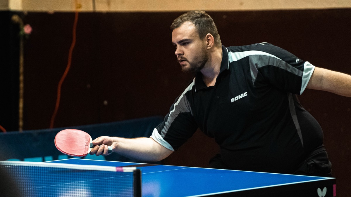
Table tennis gives a receiver a fraction of a second, which means the good players are not reacting to the ball so much as predicting it. Where that prediction happens in the brain has produced inconsistent imaging results. So experts and non-experts watched serve videos cut off at the moment of racket-ball contact and guessed the trajectory, with scanning throughout and a pattern-based analysis rather than a region-by-region one.
The brain patterns separate experts from non-experts at about 90 percent. distinguished 29 table tennis experts from 34 non-experts with 90.48 percent accuracy. The regions contributing most to that separation were the left , the vermis, the right middle temporal pole, the inferior parietal cortex, the bilateral paracentral lobule and the left .
An exceptional ability to an opponent’s action accurately is paramount for competitive athletes and highlights their experiential mastery. Action observation has conventionally been associated with specific brain regions, and neuroimaging discrepancies have persisted, which is the gap this study set out to address.
The task was built to require prediction. Participants pressed a button to predict where the ball would go in a table tennis serve video, truncated at the moment of racket-ball contact. The control was pressing any button while viewing a static image of that video’s first frame. The ball’s flight is never shown, so there is only the server’s body to read.
Functional magnetic resonance imaging data were collected from 29 table tennis experts and 34 non-experts, and both univariate analysis and multivoxel pattern analysis were applied. The second asks a different question from the first: not whether a region is more active, but whether the distributed pattern of activity can tell an expert brain from a novice one.
It could, with what the authors call moderate accuracy of 90.48 percent. The regions contributing most to the differentiation were the left cerebellum, the vermis, the right middle temporal pole, the inferior parietal cortex, the bilateral paracentral lobule and the left supplementary motor area. The authors read this as brain regions associated with cognitive conflict monitoring and motor cognition contributing to expert anticipation. Keep the measure in view: this is a classifier’s accuracy across 63 people in one sport, not a measurement of how anticipation works.
wat. A classifier told table tennis experts from novices nine times in ten, mostly from cerebellum and motor areas rather than visual ones.
wat wat
The regions that carry the difference are not visual. Cerebellum and supplementary motor area belong to motor cognition, which fits the idea that an expert reads a serve by simulating the stroke rather than by watching the ball harder.
How do we know?
Ji and colleagues collected functional magnetic resonance imaging from 29 experts and 34 non-experts predicting ball trajectory from serve videos truncated at racket-ball contact. The control was pressing any button while viewing a static first frame, and the analysis was both univariate and multivoxel.
What is still unknown
Sixty-three participants in one sport, with the key result an accuracy figure for a classifier. The authors describe 90.48 percent as moderate accuracy, and classification accuracy is not a measure of how anybody anticipates.
Learn something?
Where to next
 What do the eyes do in the instant before an accurate shot?expertise
 How much of expert performance does practice explain?expertise