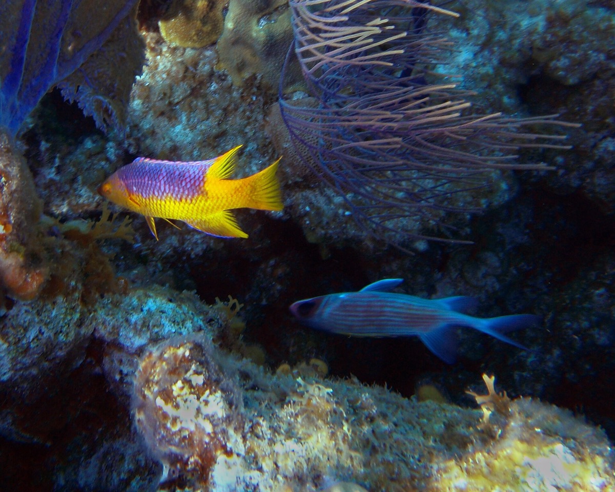
An animal changing color cannot see most of its own body, which leaves an obvious gap: nothing checks whether the change came out right. Skin that detects light has been found in many color-changing species, and its function has stayed unclear. Schweikert and colleagues worked through the morphology, physiology and optics of in the hogfish. What is the skin looking at?
Its own pigment. The light reaching the skin’s opsin is filtered by the sitting directly above it, so when pigment disperses it absorbs the short-wavelength light that opsin needs. The receptors are a morphologically specialized population nested beneath the pigment cells. One possible function is monitoring: the skin is not sampling the scene so much as watching its own color change happen.
Dynamic color change has evolved several separate times, and its physiology has repeatedly been linked to light-sensitive skin, largely through studies of excised skin preparations. Despite how widespread dermal photoreception is, both how it works and what it does for color change have remained poorly understood.
Part of the difficulty is that skin is a strange place for a light sensor. It cannot form an image, it is spread across the body, and whatever it detects is already filtered by everything lying over it. That last point turns out to be the whole story.
Schweikert and colleagues examined the morphology, physiology and optics of dermal photoreception in hogfish, Lachnolaimus maximus. They localized the skin’s SWS1 opsin to a morphologically specialized population of putative photoreceptors, and found those receptors nested beneath the chromatophores.
When chromatophore pigment disperses, it selectively absorbs the short-wavelength light that SWS1 needs to be activated. The receptor is therefore reporting, moment to moment, how much pigment is spread above it. One possible function of dermal photoreception in hogfish is to monitor the chromatophores and detect information about how the color change is performing. That gives a loop to a process which appeared to have none.
wat. The light-sensitive cells sit under the pigment, so the only thing they clearly see is whether the pigment has moved.
wat wat
It supplies the missing feedback loop in color change, turning light-sensitive skin from a puzzle into a plausible sensor for checking performance.
How do we know?
Schweikert and colleagues examined the morphology, physiology and optics of dermal photoreception in hogfish, localized the SWS1 opsin in the skin, and measured how chromatophore pigment activity alters the light transmitted to it.
What is still unknown
The authors offer monitoring as one possible function, supported by the optics and the anatomy rather than by a behavioral test showing a fish correcting its color using this signal.
Learn something?
Where to next
 A fish has no cortex like ours. So how does it tell faces apart?fish
 Why would a fish choose the turbulent water over the smooth?fish
 What decides where billions of migrating birds come down to rest?light