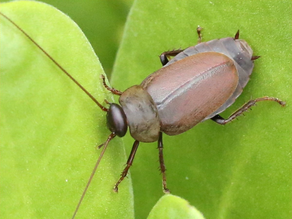
Protein crystals for X-ray work are normally grown in a laboratory from pure, uniform samples, which is a long way from any living body. A few have been found inside cells. Inside the gut of a developing embryo, in a cockroach, there is a stranger case: crystals that are food.
Milk protein crystals, from a live-bearing cockroach. The structure was solved to , 1.2 angstroms, from crystals taken out of the midgut of an embryo of Diploptera punctata, the only known cockroach. A heterogeneous, , lipid-bound protein had crystallized inside a living animal, doing its job there.
Macromolecular crystals for X-ray diffraction studies are typically grown from pure and homogeneous samples. There are, however, examples of protein crystals identified in vivo, and recent developments in micro-crystallography and X-ray free-electron lasers have allowed several structures to be determined from crystals grown inside cells.
This case goes further. The authors report an atomic resolution structure, at 1.2 angstroms, of heterogeneous milk proteins grown inside a living organism in their functional niche.
The organism is unusual in its own right. The crystals were isolated from the midgut of an embryo within Diploptera punctata, the only known viviparous cockroach, meaning the only one that bears live young and therefore has to feed them internally.
What is being described is a working biological material rather than a laboratory product. The crystal is heterogeneous, glycosylated and lipid-bound, which is everything crystallographers normally avoid, and it still yields a structure at atomic resolution.
wat. The only live-bearing cockroach feeds its embryos on protein crystals.
wat wat
A crystal that forms in a body and feeds an embryo is a reminder that the orderly structures of a laboratory also occur as biology.
How do we know?
Banerjee and colleagues isolated the in vivo grown crystals from embryo midguts and determined the structure directly, using micro-crystallography methods developed for crystals grown inside cells.
What is still unknown
One species and one crystal type, described structurally rather than tested as a nutritional source.
Learn something?